
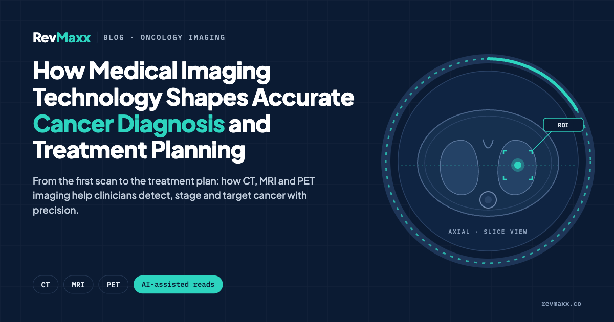
Cancer diagnosis has evolved from a process heavily reliant on invasive procedures to one where sophisticated imaging technology often provides the first definitive evidence of disease.
The choice of imaging modality can determine whether a tumor is detected early, whether treatment targets the right areas, and ultimately whether patients receive care that matches the specific characteristics of their cancer. For healthcare providers managing complex cases, understanding the strengths and limitations of each imaging approach has become as critical as selecting the right therapeutic protocol.
The Role of Advanced Imaging in Early Cancer Detection
Modern cancer screening programs depend on imaging technologies that can identify abnormalities before symptoms appear. Mammography remains the standard for breast cancer screening in average-risk populations, typically recommended annually or biennially for women over 40. Digital mammography detects microcalcifications and masses, but its effectiveness decreases in women with dense breast tissue, where normal glandular tissue can obscure tumors on X-ray images.
For patients at elevated risk—those with BRCA mutations, strong family histories, or previous chest radiation—supplemental screening with magnetic resonance imaging offers substantially higher sensitivity.
MRI uses powerful magnets and radio waves to create detailed cross-sectional images, distinguishing between different tissue types based on their water content and biochemical properties. This technology excels at detecting small invasive cancers and can identify bilateral disease that single-view imaging might miss.
When Standard Screening Requires Enhanced Visualization
Certain clinical scenarios demand imaging that goes beyond routine screening capabilities. Women with extremely dense breast tissue, comprising roughly 40 percent of the screening population, face a documented increased cancer risk and reduced mammographic sensitivity.
In these cases, centers with comprehensive breast health programs may incorporate advanced imaging protocols that layer multiple technologies to improve diagnostic accuracy.
Cleveland Clinic Abu Dhabi’s Breast Health & Cancer Program utilizes breast mri as part of its comprehensive diagnostic approach for high-risk patients, combining this with 3D mammography and image-guided biopsy services to create a complete picture of breast health. The program addresses the full spectrum of breast conditions, from screening through treatment and reconstruction, serving women throughout Abu Dhabi who require specialized breast care services.
Distinguishing Between Screening and Diagnostic Imaging
Screening imaging looks at asymptomatic patients to find disease early. Diagnostic imaging investigates a known problem—a palpable lump, nipple discharge, or an abnormality found during screening. Diagnostic studies typically involve additional views, magnification, ultrasound correlation, or contrast-enhanced techniques. The diagnostic pathway often requires multiple imaging sessions before arriving at a definitive recommendation for biopsy or continued surveillance.
Imaging’s Critical Role in Treatment Planning
Once cancer is confirmed through biopsy, imaging determines the extent of disease and guides treatment decisions. Staging scans assess whether cancer has spread beyond the primary site to lymph nodes or distant organs. CT scans evaluate the chest and abdomen, bone scans detect skeletal metastases, and PET scans highlight metabolically active tumor deposits throughout the body.
For breast cancer specifically, pre-treatment MRI can reveal additional tumor foci in the same breast or the opposite breast that weren’t apparent on mammography. This information directly affects surgical planning—whether a patient needs lumpectomy or mastectomy, whether both breasts require surgery, and how much tissue must be removed to achieve clear margins. However, MRI also detects benign findings that require additional workup, which can delay treatment.
The decision to use pre-operative MRI involves balancing thoroughness against the anxiety and logistics of extended evaluation.
Documentation Standards for Imaging in Cancer Care
Accurate medical coding ensures that imaging studies are properly documented for reimbursement, quality tracking, and clinical research. The complexity of cancer diagnosis requires precise classification of procedures and findings. Healthcare systems rely on standardized coding frameworks to capture the type of imaging performed, the anatomical area examined, and whether contrast material was used. Revenue cycle management platforms increasingly provide resources to help medical coders navigate these classification systems, with detailed guidance available through platforms like ICD-10-CM coding references that clarify documentation requirements for diagnostic procedures.
Proper coding matters beyond billing. It creates data that tracks patterns in imaging utilization, identifies variations in practice, and supports research into which imaging strategies yield the best outcomes. When imaging studies are coded incorrectly, the entire downstream data chain suffers, affecting everything from quality metrics to clinical trial enrollment.
Monitoring Response and Detecting Recurrence
During active treatment, imaging monitors how tumors respond to therapy. Shrinking masses indicate effective treatment, while growing or new lesions suggest resistance and the need for a different approach. Oncologists use standardized criteria to measure tumor dimensions across serial scans, calculating response rates that inform whether to continue, modify, or abandon a particular treatment regimen.
After treatment completion, surveillance imaging watches for recurrence. The intensity and duration of follow-up depends on cancer type, stage at diagnosis, and treatment received. Breast cancer survivors typically receive annual mammograms indefinitely, while other imaging is reserved for patients with symptoms or those at particularly high risk for distant recurrence. The challenge lies in detecting recurrence early enough to treat effectively while avoiding excessive imaging that exposes patients to radiation and generates false alarms requiring invasive workup.
Balancing Imaging Benefits Against Risks
Every imaging decision involves tradeoffs. CT scans deliver radiation exposure that accumulates over repeated studies, potentially increasing cancer risk decades later—an especially important consideration for young patients requiring long-term surveillance. MRI avoids ionizing radiation but requires patients to lie still inside a confined space for 30 to 60 minutes, which many find intolerable. Contrast agents used to enhance image quality can cause allergic reactions or kidney damage in vulnerable patients.
Cost represents another constraint. Advanced imaging is expensive, and unnecessary studies strain healthcare budgets while subjecting patients to procedures they don’t need. Evidence-based guidelines help clinicians order appropriate imaging at appropriate intervals, but guidelines can’t address every clinical scenario. Experienced oncologists weigh the probability of finding actionable information against the burden of testing, individualizing surveillance based on each patient’s specific risk profile.
Integration of Imaging with Molecular and Genetic Information
Cancer care increasingly combines anatomical imaging with molecular diagnostics. Genomic testing of tumor tissue reveals mutations that predict treatment response, while liquid biopsies detect circulating tumor DNA that may signal recurrence before imaging shows visible disease. These approaches complement rather than replace imaging. A rising tumor marker or positive circulating DNA prompts imaging to locate where disease is growing. Conversely, an imaging abnormality requires tissue sampling for molecular characterization that guides targeted therapy selection.
Treatment options continue to expand based on tumor biology rather than just location and stage. Detailed information about breast cancer treatment approaches reflects how therapy selection now incorporates hormone receptor status, HER2 expression, and genomic risk scores alongside traditional staging. Imaging remains essential for measuring anatomical response, but it works within a broader framework of biological understanding that shapes modern oncology practice.
Looking Forward in Cancer Imaging
Artificial intelligence applications are beginning to assist radiologists in detecting subtle abnormalities and predicting tumor behavior from imaging features. Machine learning algorithms trained on thousands of scans can highlight suspicious areas for human review, potentially reducing missed cancers and variability between readers. However, these tools require validation across diverse populations before widespread clinical adoption.
The evolution of cancer imaging reflects a broader shift toward precision medicine, where diagnostic accuracy enables increasingly tailored treatment. As imaging technology advances, the fundamental challenge remains unchanged: obtaining sufficient information to guide effective treatment while minimizing burden on patients. Success requires not just sophisticated equipment but thoughtful clinical judgment about when advanced imaging truly changes management and when simpler approaches suffice.